Exploring How AI Tools Can Improve Lung Cancer Detection
For people at high risk for lung cancer, annual screening with CT (computed tomography) can help reveal areas of abnormal growth, or nodules, before they become cancerous or in early stage of disease when they are easier to treat.
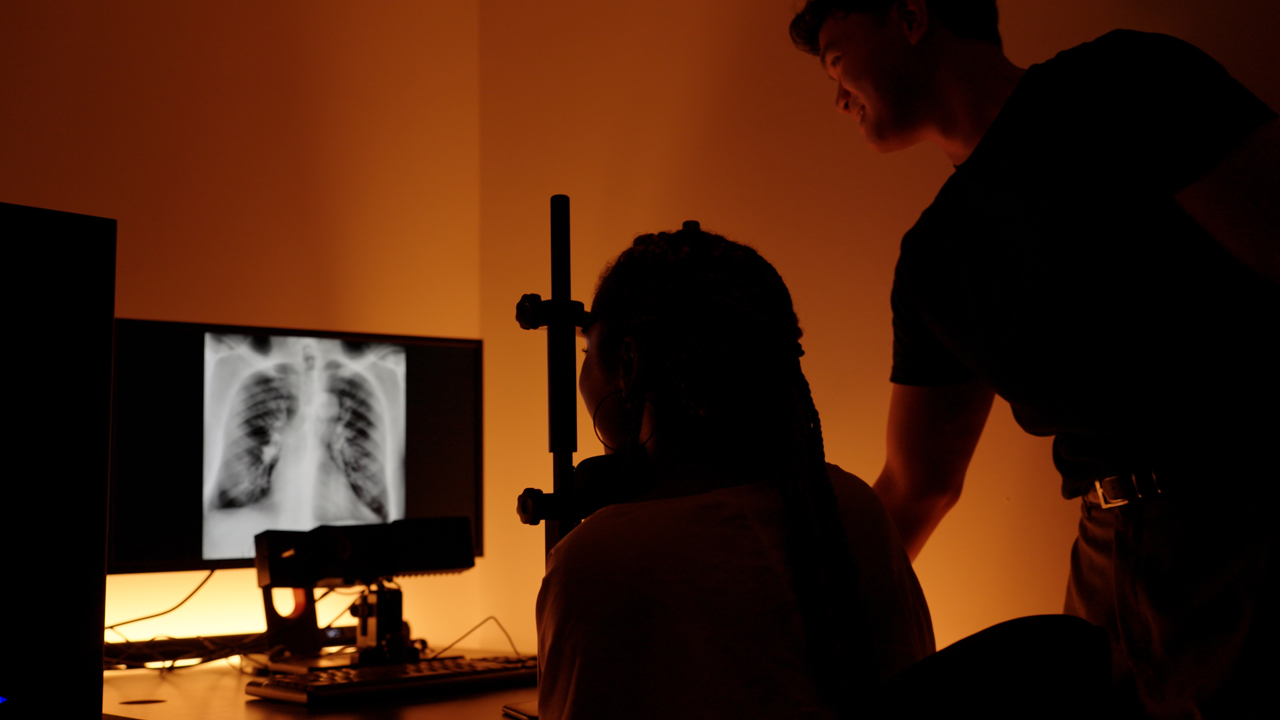
However, studies consistently show that radiologists fail to detect lung nodules in about 30 percent of abnormal CT scans. One study found that more than half of patients who were diagnosed with lung cancer had nodules in their scans the year before that were missed.
“If a radiologist misses an abnormality, you’re not treating the patient. That immediately can lead to potential patient harm. So, we care a lot about misses,” says Robert G. Alexander, Ph.D., a cognitive neuroscientist and assistant professor of psychology and counseling, who leads research examining how eye movements influence medical image perception. “And in most instances, we don’t really know exactly why a miss happens,” says Alexander.
To shed light on what the culprits are in cases of missed lung nodules, Alexander, along with Talia Lilikakis, a first-year osteopathic medical student in the life sciences/osteopathic medicine (B.S./D.O.) program in his research group, recently published a comprehensive review of research on the topic in the American Journal of Roentgenology (AJR), a leading radiology journal. The article points to three categories of factors that contribute to the problem: (1) nodule characteristics, such as size and density (smaller and less dense nodules are more frequently overlooked); (2) technical factors such as poor image quality; and (3) reader limitations, such as radiologists being tired or distracted or relying too much on artificial intelligence (AI) tools that assist in nodule detection.
With these findings in hand, Alexander says that researchers can design experiments to determine the extent to which each factor prevents radiologists from spotting nodules and how to help overcome that challenge.
Alexander and his student team, called the Human Factors and Neuroscience (HFAN) research group, are right in the mix with their work. They have embarked on a project that applies their signature interdisciplinary approach—combining psychology, cognitive science, and AI research—to assess how AI tools can most effectively alert radiologists of the possible presence of a nodule. The ultimate goal of the research team is to integrate AI tools with human expertise and clinical workflow to improve healthcare. Although radiologists have no control over some of the factors that confound nodule detection—particularly the characteristics of the nodule—Alexander sees the potential for technical advances in imaging and AI processing to make even nodules that are very small, less dense, or obscured by surrounding anatomy more apparent. “We actually don’t rule out the possibility of improvement in any of these domains,” he says.
Are AI Tools a Help or Hindrance?
As researchers get a handle on the factors that thwart lung nodule detection, Alexander says that there are pressing questions about the circumstances in which AI tools may help flag possible nodules, and how they can best convey information to radiologists.
The AJR article provides a lay of the land: Whereas some studies report AI tools boosting radiologists’ ability to detect nodules (in one study by 24 percent) and reducing the amount of time spent interpreting images, particularly when the tools were used as assistants working alongside radiologists, others did not see improvements. Studies have also found AI tools were less sensitive at spotting nodules with lower density and poorly defined margins—the same nodule types that are more likely to evade radiologists.
Even more concerning is the potential for the technology to exacerbate reader-related limitations. As Alexander and the other authors of the AJR article point out, AI tools can give false positive results, detecting possible nodules that are not actually concerning areas. And radiologists are sometimes biased toward agreeing with the AI tools, which can lead to unnecessary monitoring and fear for the patient.
“Some of the tools are quite good and useful, and it’s more a question of useful in what way, and what is the right way to approach them,” Alexander says.
To delve into features of AI tools that lend them greater utility, Alexander and his HFAN lab are measuring how quickly radiologists find lung nodules in CT and X-ray images when a tool provides a general cue (“find the nodule”) compared with a precise cue with information about the nodule (“find the lobulated nodule”). The team has conducted experiments with about 50 radiologists so far, and is about one year into the four-year project, which is funded by a National Institutes of Health (NIH) grant.
Alexander notes that commercial AI tools vary a lot in the cues they provide radiologists, including about the shape and location of potential nodules. And findings are mixed about the impact of these cues. Although Alexander and his team expect that precise cues would help radiologists zero in on concerning areas faster and more efficiently (with fewer eye movements), the additional information could have unintended consequences. Attention may instead be drawn too narrowly on one suspected finding, causing radiologists to overlook other abnormalities elsewhere in the image. The current project is the first to quantify the effect of these cues on radiologists’ eye movements as they interpret medical images. The approach will also allow the team to analyze how radiologists review CT images—where they look and how they scroll through the images as they inspect them—and whether individual search strategies are impacted differently by precise cues. Discoveries from this work can inform how future AI systems are designed.
From Classroom to Research Leadership
Alexander was drawn to New York Tech because of the opportunities he has to teach and mentor. Alexander’s commitment to undergraduate research mentorship was recently recognized with New York Tech’s Presidential Excellence Award for Student Engagement in Research, Scholarship, or Creative Activities at the 2026 faculty and staff convocation.
Teaching also allows Alexander to involve students from a range of disciplines in research, from psychology to computer science and finance.
Such was the case for Lilikakis, the B.S./D.O. student in his research group. Lilikakis hadn’t planned to do undergrad research but became enthralled by the introduction to psychology course that Alexander taught in her freshman year—and his own research that he described. She couldn’t believe it when Alexander invited students in the class—and noticing both her enthusiasm and exceptional performance in the course, Lilikakis personally—to participate in the research.
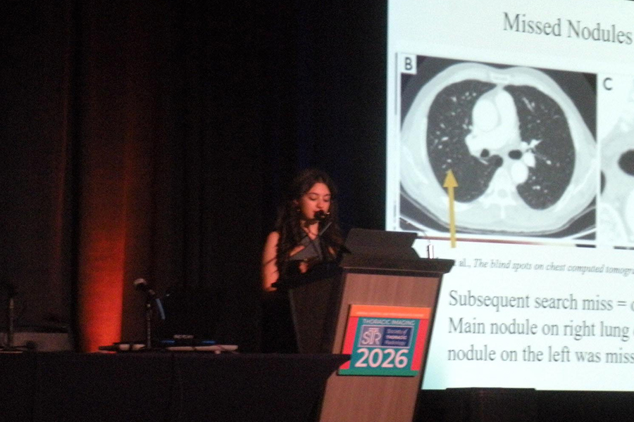
Over the last three years, Lilikakis, like other students in Alexander’s group, has developed a range of skills, from learning experimental basics to designing her own experiments, analyzing data, and mentoring newer members in the HFAN group. Lilikakis learned from Alexander how to perform a literature review and collaborated with radiologists to put together the AJR article, for which she is the second author. Lilikakis presented its findings at the 2026 Annual Meeting of the Society of Thoracic Radiology (STR), winning an award for Best Student Oral Scientific Presentation.
Through a New York Tech Teaching and Learning with Technology (TLT) grant awarded to Alexander, Lilikakis also helped conduct additional research at the STR conference, examining how radiologists are trained to interpret individual medical images and what strategies experienced radiologists now teach to their own trainees.
As Lilikakis works to complete her D.O., she plans to focus on neurology and fully intends to make research an ongoing part of her career. In the immediate term, she will continue to work on the project to understand the impact of precise cues from AI tools. She and other members of the HFAN lab recruited radiologists for the study and carried out the first set of experiments at the STR meeting.
Lilikakis hopes that some of her future research will also focus on AI. “As AI becomes more and more prevalent in everything that we do, and especially in medicine, I feel like it has the potential to be very helpful if it’s used correctly. And so, while it’s growing, I think proper research needs to be done now,” she says.
“I’m very grateful that, as an undergrad, I got research experience. That is insane and very difficult to do. And then to be able to write my own papers and present at conferences, and to do that with Dr. Alexander’s mentorship, is so valuable to me,” Lilikakis says.
As AI becomes more and more prevalent in everything that we do, and especially in medicine, I feel like it has the potential to be very helpful if it’s used correctly.
-Talia Lilikakis
More News
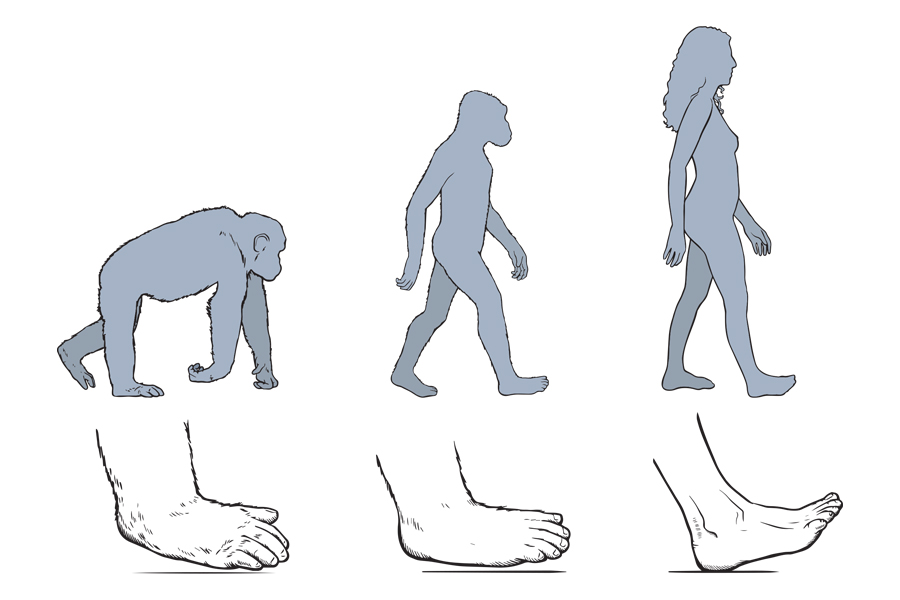
The Evolutionary Significance of Humans’ Unique Heel-Toe Walking Pattern
Associate Professor of Anatomy Nathan E. Thompson, Ph.D., contributes to research unveiling clues to the origin of humans’ stride pattern.
Robust Research
New York Tech’s commitment to research paves the way for important and groundbreaking work. Meet three Ph.D. students whose work may improve disaster response, recovery after muscle and bone injuries, and anticancer therapies.
NYITCOM-Arkansas’ Rajagopalan Receives $573K NIH Grant for Cardiovascular Disease Research
The funding will further Associate Professor Viswanathan Rajagopalan’s research regarding the impact thyroid dysfunction has on cardiovascular disease.

NYITCOM Student Receives American Heart Association Scholarship
A student researcher from the College of Osteopathic Medicine (NYITCOM) has secured the prestigious Student Scholarship in Cardiovascular Disease from the American Heart Association.

Ph.D. Student Uses Game Theory to Model Manhattan
Electrical and computer engineering Ph.D. student Helen Wu uses game theory to study the impact of congestion pricing in New York City.

Fellows’ Field Notes: Bofan He and Christine Lee
Edward Guiliano Global Fellowship recipients Bofan He and Christine Lee reflect on their experiences as they traveled to conduct academic research.