Keeping Science on Its Toes
In the late 1800s, researchers discovered, in separate studies, that horses and humans have a small bone at the tip of their hoof and finger, respectively. Since then, research on the anatomical development of many animals—including horses, humans, and other species—has forged on, but little attention has since been given to this bony structure, which went unnamed for more than 100 years.
Now, College of Osteopathic Medicine (NYITCOM) researchers, including Professor of Anatomy Nikos Solounias, Ph.D., and medical student Shannon Smith, are resurfacing this small but mighty structure and ensuring that it does not become lost within scientific literature. In a study published on January 10 by PeerJ, they make the case for why this structure—which they dub “the bony cap”—may play a large role in digit (finger and toe) regeneration and nail growth.

The researchers analyzed the anatomy of fetal and adult humans, horses, and cats via microscopic tissue samples and scans obtained through micro-computed tomography (micro-CT), an imaging technique that captures microscopic images of specimens’ internal spaces and recreates virtual 3-D models without dissection.
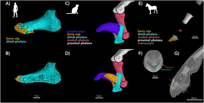
They discovered that the bony cap was present on all digits in all three species. During development, the bony cap is a distinct anatomical structure that later fuses with the area at the tip of the digit called the distal phalanx.
The project began when Solounias first noticed the presence of the bony cap in developing humans, which he notes was a good place to start, as human anatomy can be much simpler than other species. An analysis of adult bones from the NYITCOM Anatomy Lab showed the presence of a bony cap that had fused with the distal phalanx.
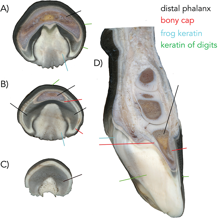
The study was extended to horses, which have very complex anatomy because their hooves contain five separate digits fused into one, as Solounias noted in a pioneering 2018 study. Much like humans, horses had a very distinct bony cap during development that seemed to later fuse with the distal phalanx by adulthood.
Cats, however, proved to be a particularly interesting case. Unlike the bony caps of horses and humans, those in cats were hollow and adjacent to the distal phalanx. The authors believe that these distinct features may play a role in allowing cats to retract their claws.
Regardless of these differences, the researchers conclude that “the bony cap may play a significant role in nail formation in humans, claw formation in cats, and hoof formation in horses.” They also call for future studies that examine how the bony cap may impact nail formation in other animals, stating, “Its ubiquity across mammals, and even tetrapods (four-footed animals), needs further consideration.”
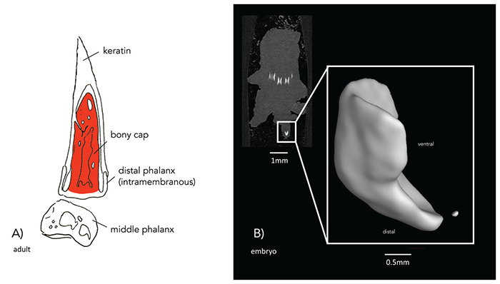
In addition, the findings may improve the scientific understanding of digit regeneration. For example, young babies re-heal and grow back the distal phalanx, but this regeneration likely requires the bony cap to act as a catalyst. To further demonstrate this, the authors cite existing animal studies that found amputated digits closer to the limb were unable to regenerate bone.
“When considering this, it may be possible that the bony cap is responsible for bone regrowth in amniotes (mammals, birds, and reptiles),” the authors write.
The study is the result of a collaboration between NYITCOM faculty, students, and staff. Smith assisted Solounias with his research and co-authored the paper, while Research Technician Kelsi Hurdle captured high-resolution images of embryonic specimens via the micro-CT scanner housed in the Department of Anatomy’s Visualization Center.
Smith and Solounias also collaborated externally with comparative and embryology expert Laurel Yohe, Ph.D., a researcher affiliated with the University of North Carolina at Charlotte and Yale University. Yohe facilitated the data collection and scanning of multiple adult specimens via facilities at Yale University and co-authored the study. She also helped in writing the manuscript.
More Features
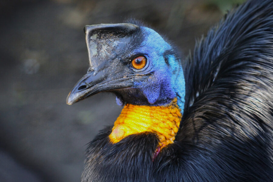
The Cassowary’s Impact on the Rainforest
NYITCOM-Arkansas Assistant Professor Todd Green, Ph.D., sheds light on a bird species that plays an impactful role in rainforest conservation.

NYITCOM Student is Congresswoman’s Guest at State of the Union
College of Osteopathic Medicine (NYITCOM) student Briteny Xu was Congresswoman Grace Meng’s guest at the State of the Union address in Washington, D.C., where they advocated for the Affordable Connectivity Program.

Baking Her Way to a Business Degree
Student Michelle Charidemou is studying for her bachelor’s degree in business administration to help take her bakery to the next level.

New York Tech Joins Engineering Doctoral Consortium
New York Tech is one of nine New York City-area universities in a new consortium through which engineering doctoral student can take courses at each other’s institutions without any additional tuition, as part of a new multi-school agreement.

Celebrating 15 Years of FRIENDS
On October 25, the School of Architecture and Design celebrated its 15th Annual Alumni and FRIENDS Reception, raising a record-breaking $401,300 in support of student scholarships, new initiatives, capital improvements, travel, and lectures.

Guiliano Global Fellows: Studying Grip Strength in Mongolia
Throughout each year, a handful of New York Tech students are selected to complete academic research projects at least 200 miles away from their campus through the Edward Guiliano Global Fellowship Program.